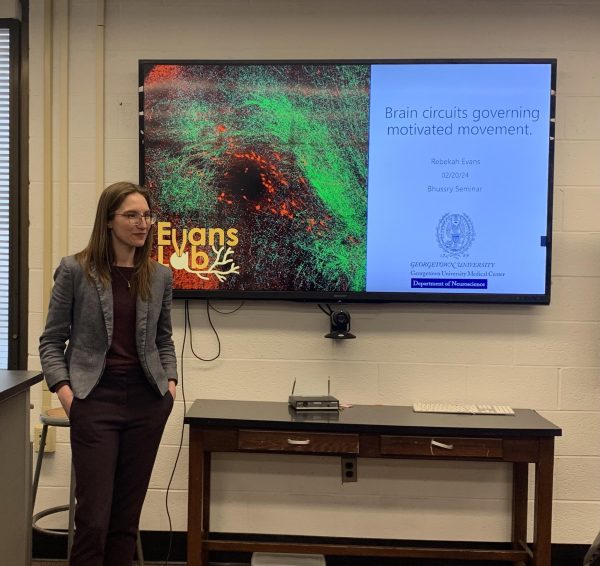
A Georgetown University neuroscience professor discussed her research on motor pathways at the Georgetown University Medical Center on Feb. 20.
The department of biochemistry hosted the talk on neuroscience research as part of its Bhussry Seminar series, weekly research talks hosting scientists from various institutions including Georgetown. The event featured Rebekah Evans, assistant professor in the department of neuroscience, who presented her work on motivated behaviors.
Evans’ research focuses on a motor pathway that originates in the basal ganglia, a part of the brain involved in voluntary movements, learning and motivated behaviors. In addition, the basal ganglia is associated with Parkinson’s disease. Evans’ work concerns the substantia nigra pars reticulata (SNr) and globus pallidus externus (GPe) — two parts of the basal ganglia — and their interactions with a region in the brainstem called the pedunculopontine nucleus (PPN), which is also involved in movement and is impaired in Parkinson’s patients.
According to Evans, the SNr and GPe both inhibit the PPN by releasing an inhibitory neurotransmitter, a chemical signal between neurons. Evans said her research focuses on the mechanisms underlying these two inhibitory connections.
“‘Why does it have these two output pathways that can both modulate movement but through completely different mechanisms?’” Evans said at the event. “Well, we wanted to understand a little bit about how that was working.”
To begin to understand the SNr and GPe connection to the PPN, Evans’ team injected mice with a protein that activates neurons. Evans hypothesized, based on existing literature, that stimulating the SNr would inhibit movement while stimulating the GPe would increase movement — but found the opposite. The SNr increased movement while the GPe inhibited movement.
“It’s not our second hypothesis, that they might do the same thing because they’re both inhibiting the same structure,” Evans said. “They’re actually doing opposite things, even though they’re both causing inhibition.”
Evans then investigated the manner in which the SNr and GPe interact with the brainstem to carry out opposite movement behaviors.
“So we want to ask two questions,” Evans said. “One is, ‘Do these two structures of the basal ganglia inhibit different parts of the PPN anatomically? Or do they inhibit different cell types that are molecularly defined there?’”
To further investigate the connections between these brain regions, Evans’ team performed electrophysiology experiments, stimulating mouse brains with light then measuring inhibitory currents to three types of neurons in the brainstem.
Evans found that the SNr and the GPe connect to different neurons in different regions of the brainstem. The SNr connects to two types of neurons called cholinergic and glutamatergic neurons in both the front and back of the PPN, while the GPe connects to only glutamatergic neurons in the back of the PPN.
“The GPe connecting only to the glutamatergic neurons is really interesting,” Evans wrote to The Hoya. “We don’t know the full extent of its significance yet, but think that this output pathway from the GPe to the brainstem is a way for the GPe to directly modulate motor activity.”
Evans’ team found that a certain set of neurons, known as glutamatergic neurons, were vulnerable to both SNr and GPe inhibition. Evans said her team aims to work on distinguishing the differences between those neurons’ interactions with the SNr and the GPe.
“We don’t know for sure if these are inhibiting motion or enhancing motions to begin with,” Evans said. “So that’s one of our next steps as well.”
Evans said that teasing out the connections between the basal ganglia and the brainstem is a stepping stone to improving deep brain stimulation, which can help alleviate some symptoms in Parkinson’s patients.
“The PPN is sometimes targeted for a Parkinson’s Disease therapy called Deep Brain Stimulation,” Evans wrote. “If we understand the connections between the basal ganglia and the PPN, we can develop more precise stimulation locations and stimulation patterns to optimize Deep Brain Stimulation treatment.”
Stella Alimperti, an associate professor in the department of biochemistry and molecular and cellular biology at Georgetown’s School of Medicine, attended Evans’ talk and said that Evans’ work on motor pathways will have implications for furthering the understanding of neurological diseases.
“The most important part is really to figure out the key molecules that are involved in diseases, like neuro diseases such as Parkinson’s disease, and really the novel methods that she is trying to develop and use,” Alimperti told The Hoya.