On Jan. 16, the Georgetown University Medical Center announced the publication of a study that showed the reversal of amnesia following repetitive head injury in mice.
Mark Burns, a professor of neuroscience and the director of the Laboratory for Brain Injury and Dementia, led the research, published in the Journal of Neuroscience. The lab’s findings suggest the brain can reset to its normal function after head trauma, leading to less memory loss.
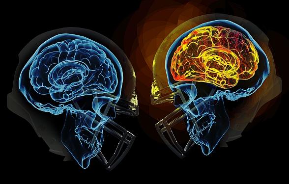
The study, which was prompted by over a decade of research on chronic traumatic encephalopathy in football, used an advanced brain injury model, known as controlled cortical impact, to inflict brain injury onto the mice proportional to what young football players face on a weekly basis.
“Ours was really one of the first animal models that took a large number of head impacts and we spread them out over a long period of time, and then we compressed them into short periods of time,” Burns told The Hoya.
The researchers then trained a group of mice to associate a mildly uncomfortable shock with a specific environment. This process, generally known as conditioning, is common in animal memory studies.
Isaac Cervantes-Sandoval, professor of biology, uses this conditioning technique for his own research, which focuses on the neurobiology of memory formation and forgetting.
According to Cervantes-Sandoval, conditioning allows for researchers to study learning and memory patterns in animals.
“The animals form the association between the environments or the cage where they are, and then the punishment,” Cervantes-Sandoval told The Hoya. “So when they’re exposed back to the same place they remember that they were shocked at that place, they react again with the freezing behavior. ”
The Burns Research Group used conditioning to assess the mice’s memory capabilities after the initial head injury. While the animals were mildly shocked, the researchers used a biomarker to track the neurons activated during exposure. These neurons are then connected in a network known as a memory engram. An engram is a specific neural network that activates during an experience and displays activation again when exposed to the same stimulus, leading to their focus in memory studies.
The researchers also used optogenetics, using a laser to reactivate the memory engram in the mice that underwent memory loss.
“When we look in the hippocampus, any neuron that’s yellow is an engram neuron. So that was involved in learning the memory or was active at the time of learning the memory,” said Burns. “That’s also the neuronal network that we’re turning back on again with the optogenetic implant.”
The mice that faced brain injury once again associated the environment with the stimulation, as they showed the freezing behavior they demonstrated before head injury. This showed the researchers that the mice regained the association memory between the environment and the stimulus.
While these findings spark optimism that memory loss can be halted, Daniel Chapman, an M.D./Ph.D. candidate in the lab and an author of the research paper, highlights the limitations of the study and reversal technique.
“We have to address the fact that this used a very invasive technique for the stimulation of the brain,” Chapman told The Hoya.
The researchers are looking forward to exploring less invasive techniques of brain stimulation in humans, many of which are already used to treat other neurological diseases.
“The things that are kind of emerging in that space are things like transcranial magnetic stimulation, transcranial direct current stimulation and there are a couple other different ways to stimulate without actually needing surgery,” Chapman said.
For now, the study serves better as proof that memory loss from traumatic brain injury is not due to the death of neurons, but some other mechanism.
According to Chapman, the findings are surprising because they indicate information can be lost within the brain, but still present.
“The ability to reverse the memory is also very important in our understanding of why the memory was lost in the first place, because that suggests that the content of the information is still within the brain. It’s just locked in some kind of way,” Chapman said.
Looking forward, this group of researchers hopes to gain a greater understanding of the brain’s response to injury and are optimistic about the surplus of ongoing neuroscience developments.
“We have a lot to learn about how the brain changes or adapts to exposure to very mild, repetitive head impact.” Burns said. “It is an exciting time, and I think we’ll definitely be making progress in the next five to ten years.”

